| E-mail ID : info@iamg.in |
| E-mail ID : info@iamg.in |
Online Submission |
| Click Here For Online Submission |
| Instructions for authors |
Genetic Clinics |
| Editorial board |
Get Our Newsletter |
| Subscribe |
Send Your Feedback |
| Feedback Form |
About Us |
| IAMG |
Clinical Vignette
|
Features |
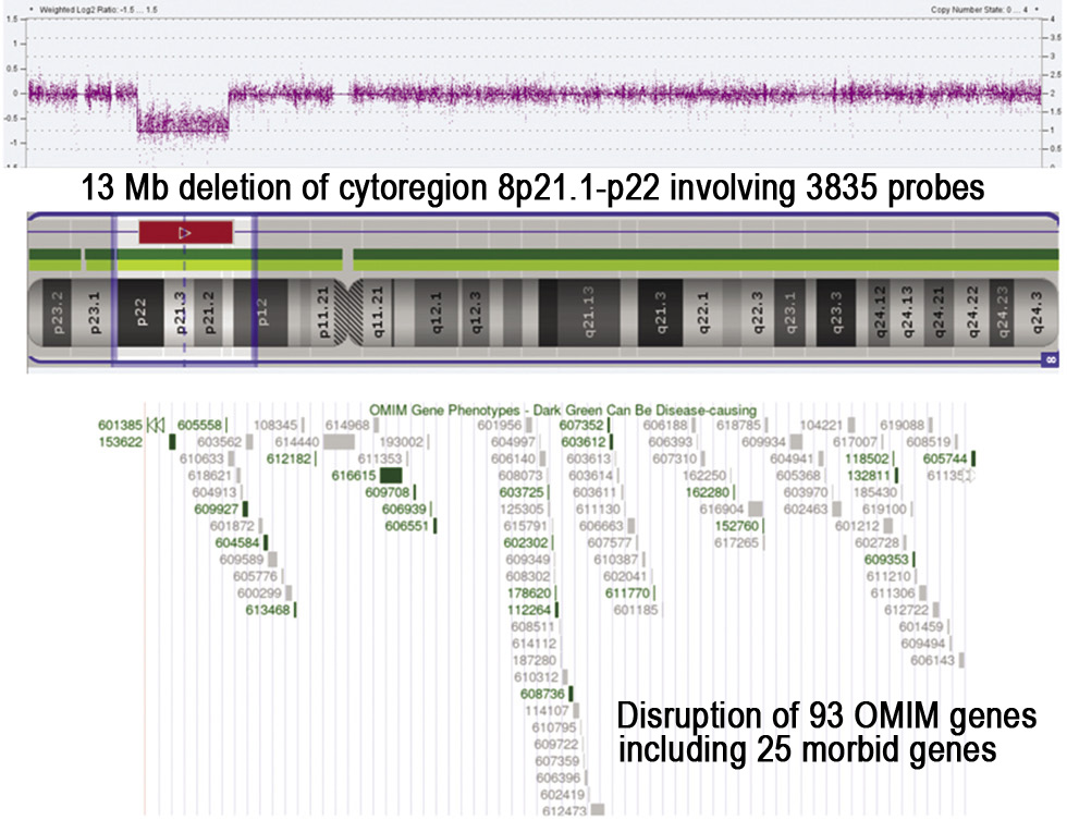
Our 13 MB chr8: |
(Izumi 3.6 MB CNV loss chr8: (GRCh38) |
(Piovani 1 MB CNV chr8: (GRCh38) |
(LaBran 11.4 MB chr8: |
ClinVar: 16.7MB Chr8: |
DECIPHER: (396045) 14.59 MB CNV loss 12798120-27386566 (GRCh38)
|
DECIPHER: 7. 15 MB chr8:8: |
DECIPHER: (327931) 3.95 MB chr8:
|
|
Sex |
Male |
Male |
Male |
Male |
NA |
Female |
Male |
Female |
|
Global developmental delay |
Yes |
Yes |
Yes |
Yes |
Yes |
Yes |
NA | NA |
|
Intellectual disability |
Yes |
Yes |
Yes |
Yes |
Yes |
Yes |
Yes |
Yes |
|
Short stature |
Yes |
No |
No |
No |
No |
Yes |
NA |
NA |
|
Microcephaly |
Yes |
No, |
No |
No |
Yes |
Brachycephaly | NA | NA |
|
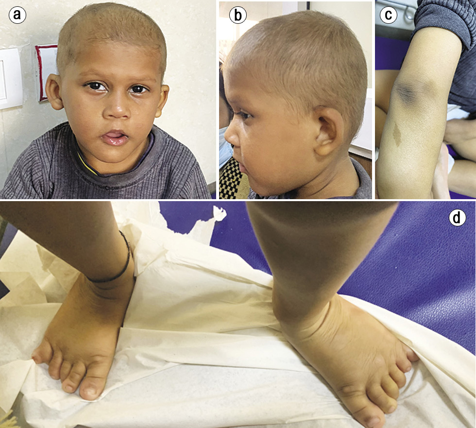
Dysmorphic features |
Hypotelorism, upslanting palpebral fissures, deep-set eyes, low-set ears, cup-shaped pinnae, long and smooth philtrum and downturned corners of the mouth. |
Deep-set eyes, mild synophrys, horizontal eyebrows, prognathism, high palate and broad uvula |
Hypertelorism, down- small
|
Deep-set eyes, mild ptosis of left eye, prominent ears, flat philtrum, thin upper lip, pointed chin |
Abnormal facial shape |
Hypertelorism, upslanting palpebral fissures, round face, thin upper vermilion, wide intermammillary distance, posteriorly rotated ears |
Oligodontia |
Upslanting palpebral fissures, prominent nasal bridge |
|
Sparse and thin hair and eyebrows |
Yes |
Yes |
Yes |
Yes |
NA |
Yes |
NA |
Yes |
|
Extremities/Joints hypermobility |
Hypotonia, bilateral post-axial polydactyly in feet |
Cubitus valgus |
Arachnodactly, valgus knee and only one groove of the right hand with distal ligamentous laxity |
Bilateral neuromuscular equinus |
Generalized hypotonia |
NA |
NA |
Hypotonia, pes cavus |
|
Unilateral renal agenesis |
NA |
NA |
NA |
NA |
NA |
NA |
Yes | NA |
|
Cryptorchidism |
No | NA | NA |
Glandular hypospadias |
Yes |
NA | NA | NA |
|
Skin pigmentary abnormalities |
No | NA | NA | Yes | Yes | NA |
NA |
NA |
|
Radiology (X ray/MRI brain) Imaging |
Brain MRI: Normal |
X ray: slipped capital femoral epiphysis and secondary avascular necrosis. Brain MRI: prominent lateral ventricles
|
Brain MRI: hyper- intensity in the long TR sequences involving posteriorly the periventricular white matter |
NA | NA |
NA |
NA |
NA |
|
Number of OMIM genes |
93 OMIM genes |
39 OMIM genes, 8 morbid genes |
1 OMIM gene |
53 OMIM genes, 11 morbid genes |
98 OMIM genes |
80 OMIM genes |
58 OMIM genes |
39 OMIM genes |
|
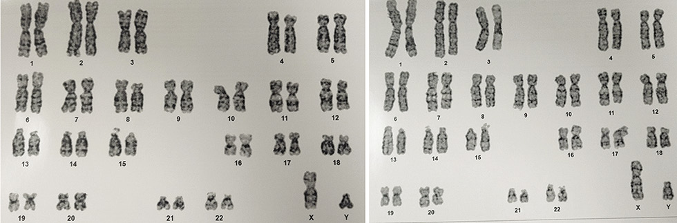
ACMG Classification |
Pathogenic ACMG CNV score: 1.55 |
Pathogenic |
Pathogenic |
Pathogenic |
Pathogenic |
Likely Pathogenic |
Pathogenic |
Pathogenic |
References
1. Hand M, et al. Mild phenotype in a patient with mosaic del (8p)/inv dup del (8p).Am J Med Genet A. 2010;152:2827-2831.
2. Izumi K, et al. 8p21 microdeletion in a patient with intellectual disability and behavioral abnormalities. Am J Med Genet A. 2011;155:3148-3152.
3. LaBranche JT, et al. 8p23. 2p22 deletion: a case report of a large deletion encompassing 8p23. 1 with additional clinical features. Clin Dysmorphol. 2020;29:207-209.
4. Orye E, Craen M. A new chromosome deletion syndrome. Report of a patient with a 46, XY, 8p chromosome constitution. Clin Genet. 1976;9:289-301.
5. Piovani G, et al. De novo 1Mb interstitial deletion of 8p22 in a patient with slight mental retardation and speech delay. Mol Cytogenet. 2014;7:1-5.
6. Pramparo T, et al. Inverted duplications: how many of them are mosaic? Eur J Hum Genet. 2004;12:713-717.
7. Riggs ER, et al. Technical standards for the interpretation and reporting of constitutional copy-number variants: a joint consensus recommendation of the American College of Medical Genetics and Genomics (ACMG) and the Clinical Genome Resource (ClinGen). Genet Med. 2020; 22: 245-257.
8. Soler A, et al. Fetoplacental discrepancy involving structural abnormalities of chromosome 8 detected by prenatal diagnosis. Prenat Diagn. 2003;23:319-322.
9. Tabares-Seisdedos R, Rubenstein JL. Chromosome 8p as a potential hub for developmental neuropsychiatric disorders: implications for schizophrenia, autism and cancer. Mol Psychiatry. 2009; 14:563-589.
| Abstract | Download PDF |