A Fetus with Trisomy 12p: Prenatal and Postnatal Presentation
Seema Thakur1, Chanchal Singh2, Manju Gupta3 1Genetic Medicine Unit, Fortis Hospital, Delhi- NCR, India 2Fetal Medicine Unit, Madhukar Rainbow Children Hospital, Delhi, India 3Department of Obstetrics & Gynaecology, Jaypee Hospital, Noida, India Correspondence to: Dr Seema ThakurEmail:seematranjan@gmail.com
1 Abstract
Trisomy of the short arm of chromosome 12 is a rare chromosomal abnormality. We have compared the ultrasound
features and autopsy features of a fetus with trisomy 12p with a previous reported antenatal case and Pallister Killian
syndrome. Ours is the second case report on fetal features of trisomy 12p.
2 Introduction
Trisomy of the short arm of chromosome 12 is a rare chromosomal abnormality with an estimated incidence of 1 per
50,000 births and only over 30 cases reported worldwide (Segel et al.,2006).The first case with trisomy 12p was reported
by Uchida and Lin (1973) due to a malsegregation of a balanced parental chromosome rearrangement (Uchida &Lin,
1973). Trisomy 12p syndrome is associated with moderate to severe psychomotor retardation, generalized hypotonia and
facial dysmorphism characterized by a round face with prominent cheeks, prominent forehead, broad nasal bridge, short
upturned nose, long philtrum, thin upper lip, broad everted lower lip, and abnormal ears (Rauch et al.,1996; Tekin et
al.,2001; Tsai et al., 2005).
We report here a 23-24 weeks fetus with trisomy 12p and compare the antenatal and postnatal features with the
previous reported fetus with 12p trisomy and Pallister Killian syndrome.
3 Case Report
A 34-year-old primigravida was referred to our fetal medicine centre in view of lower limb abnormality detected in the
anomaly scan. She had no history of fever with rash, diabetes, drug intake or radiation exposure. Nuchal translucency was
within normal range in the first trimester ultrasound. First trimester biochemistry showed low risk for Down syndrome
(PAPPA 2.27 MoM and beta hCG 0.46 MoM). Ultrasound was done using a Voluson E10 scanner (GE Healthcare,
Milwaukee, WI) equipped with a convex 4–8 MHz abdominal transducer and a 6–12 MHz endovaginal probe.
Two-dimensional ultrasound showed a single live intrauterine fetus with overall fetal growth corresponding to 20 weeks
gestation. However all long bones were below the 5th centile for gestation. There was lower limb length discrepancy. The
left femur was below the 1st centile for gestation and showed bowing. The left lower limb showed a curved tibia
and very short segment of fibula. The left foot showed valgus deformity with overcrowding of toes (Figure
1c). There was no polydactyly. Clavicle and scapula were seen. Fetal spine appeared normal. Fetal head
showed brachycephaly. There was no other structural abnormality nor any other marker for chromosomal
abnormalities. The couple was offered amniocentesis for microarray and advised consultation with paediatric
orthopaedician. However, they opted for termination of pregnancy and consented for a complete postnatal
evaluation.
On postnatal external examination, foot length was 3.5 cm corresponding to 21 weeks, crown heel length was 30 cm,
consistent with 24 weeks and crown rump length was 20.3 cm, consistent with 24 weeks and HC was 20 cm, consistent
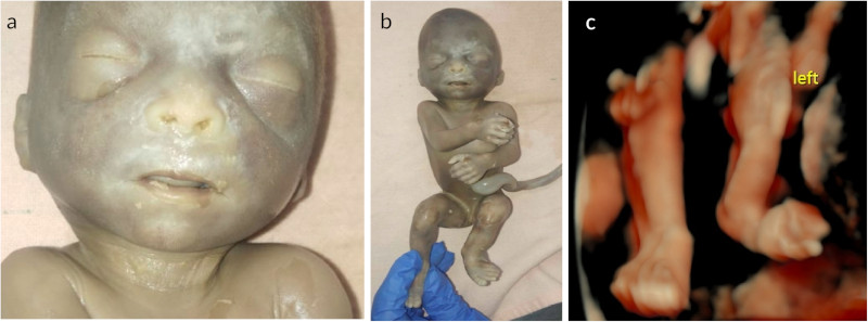
with 23 weeks gestation. There was brachycephaly, short nose, depressed nasal bridge and long philtrum (Figure 1a).
There was no cleft lip or cleft palate. Ears were low set. Neck was short. Anus was anteriorly placed. In left lower limb
there was bowing of tibia (Fig 1b). Histopathology of internal organs like liver, spleen, kidneys and lungs was
normal. Placental histopathology was normal. Histopathology of left leg showed disorganised cartilaginous
tissue.
Chromosomal microarray from fetal DNA showed a female karyotype with duplication of 18.1 MB at cytoband
12p13.33p12.1 [arr 12p13.33p12.1(803488-24653237) x 3]. This duplication has 182 genes.
Figure 1: a) Fetal autopsy evaluation showing facial dysmorphism. b) Valgus deformity and bowing of tibia
noted in the fetus. c) Antenatal ultrasound showing bowing of tibia on the left.
4 Discussion
Phenotypic similarity between trisomy 12p and tetrasomy 12p has been described in the literature. We have compared
ultrasound features of one antenatal case described previously and Pallister Killian syndrome fetus with our case in Table
1. However, it has to be kept in mind that mosaic tetrasomy 12p can have a similar pattern in chromosomal microarray as
trisomy 12p. As conventional karyotyping was not done in this case, the possibility of mosaic tetrasomy 12p could not be
entirely ruled out.
Table 1: Comparison of antenatal features of trisomy 12p and tetrasomy 12p.
Antenatalultrasonographyfeatures
Huang et al.,2012
Pallister KillianSyndrome
Fetus describedin our study
Polyhydramnios
+
+
-
Short
long
bones
+
+
+
Increased
nuchal
translucency
+
+
-
Congenital
diaphragmatic
hernia
-
+
-
Cardiac
anomaly
-
+
-
Polydactyly
-
+
-
Fetal
growth
restriction
-
Table 2: Comparison of facial features of Pallister Killian syndrome and trisomy 12p.
Features
Pallister Killiansyndrome
Huang et al.,2012
Fetus reportedin our study
Brachycephaly
+
+
+
Round
face
-
+
Coarse
facies
+
-
+, mild
Flat
facial
profile
+
+
+
Broad
nasal
bridge
+
+
+
Anteverted
nostril
+
+
+
Long
Philtrum
+
+
+
Upper
Lips
Thin
Thick
Thin
Short
Neck
+
+
+
Hung et al. reported a fetus with trisomy 12p at 30 weeks in a primigravida (Hung et al.,2012). Ultrasonography
features included polyhydramnios, short lone bones and abnormal spine curvature. Fetal facial dysmorphism included
hypertelorism, marked prenasal thickness, broad and flat nasal bridge, cleft palate, large philtrum with thickened everted
upper lip, and micrognathia.
Doray et al. stated that the three most frequent ultrasound indicators were polyhydramnios (84%), congenital
diaphragmatic hernia (CDH) (16%) and micromelia of predominantly rhizomelic type (10%) (Doray et al.,
2002).
The fetus we described also had short long bones but there was no polyhydramnios probably because of the early
gestation at detection. Left tibia was small and deformed. Histopathology of bone showed localized dysostosis.
Oligonucleotide-based aCGH showed a 35.4 MB duplication of 12p [arr 12p13.33p11.1 (0–35,400,000) × 3] in the case
reported by Hung et al. Our case had duplication of 18.1 MB at cytoband 12p13.33p12.1. Izumi et al. described a minimal
critical region for Pallister Killian Syndrome phenotype in a case with duplication of 26 genes (Izumi et al.,2014). Three
genes, ING4, CHD4, and MAGP2 represent strong candidate genes for minimal critical region of this phenotype. ING4
gene plays important role in transcriptional regulation and CHD4 gene is involved in chromatin remodelling, DNA
damage response and cell cycle control.
This case highlights the importance of a well-performed antenatal ultrasound. Down syndrome may be the commonest
chromosomal abnormality but a low risk on the combined first trimester screening does not exclude other abnormalities.
Another point to be emphasized is that any structural abnormality warrants microarray over conventional
karyotyping. A complete postnatal evaluation including infantogram and fetal autopsy is essential to confirm
ultrasound findings and to establish the diagnosis, which is instrumental in assigning appropriate recurrence
risk.
5 Conclusion
Our report further expands the spectrum of antenatal and postnatal phenotype of trisomy 12p.
References
1. Doray B, et al. Pallister-Killian syndrome: difficulties of prenatal diagnosis. Prenat Diagn 2002;
22(6):470-477.
2. Hung CC, et al. Prenatal diagnosis of a fetus with a de novo trisomy 12p by array-comparative genomic
hybridization (array-CGH). Gene 2012; 595: 178-182.
3. Izumi K, et al. Pallister–Killian syndrome. Am J Med Genet Part C Semin Med Genet 2014; 166C: 406-413.
4. Rauch A, et al. Clinical and molecular cytogenetic observations in three cases of “trisomy 12p syndrome”.
Am. J. Med. Genet 1996; 63, 243-249.
5. Segel R, et al. The natural history of trisomy 12p. Am J Med Genet 2006; 140: 695-703.
6. Tekin M, et al. De novo inverted tandem duplication of the short arm of chromosome 12 in a patient with
microblepharon. Am J Med Genet 2001; 104: 42-46.
7. Tsai AC, et al. De novo duplication of the short arm of chromosome 12: dup(12)(p13.1p13.3). Am J Med
Genet A 2005; 134A: 229-230.
8. Uchida IA et al. Identification of partial 12 trisomy by quinacrine fluorescence. J Pediatr 1973;82: 269-272.