Validation of MLPA-detected Single Exon Deletion of the DMD Gene by Multiplex PCR
Haseena Sait, Shubha R Phadke Department of Medical Genetics, Sanjay Gandhi Postgraduate Institute of Medical Sciences, Lucknow, India Correspondence to: Dr Shubha R PhadkeEmail:shubharaophadke@gmail.com
1 Abstract
Multiplex ligation-dependent probe amplification (MLPA) is the most widely used technique to detect deletions and
duplications of the DMD (dystrophin) gene that constitute two third of the cases of Duchenne (DMD)/ Becker muscular
dystrophy (BMD). However, MLPA can yield false positive results for single exon deletions owing to the presence
of single nucleotide polymorphisms (SNPs) at the ligation site. This study aimed to validate single exon
deletions detected by MLPA in DMD/BMD individuals by multiplex polymerase chain reaction (PCR).
The data of patients with DMD/BMD diagnosed by MLPA between January 2016 to February 2021 was
collated and those with apparent single exon deletions were recruited. MLPA was performed using the
P034/P035 DMD (MRC Holland) kit. Multiplex PCR was performed using Chamberlain and Beggs primer sets
with appropriate controls. Sequencing of the relevant exon of the DMD gene was planned for discordant
results between MLPA and multiplex PCR. SNPs within 8bp from ligation sites on MLPA probes and its
frequency in the South Asian population was ascertained from appropriate databases. Single exon deletion
was present in 166 (20.4%) of the affected individuals. Validation of MLPA results by multiplex PCR was
performed in 135 affected individuals for the following exons- 4, 8, 43, 44, 45, 46, 48, 50, 51, 52 and 53. We
obtained 100% concordant results for single exon deletions by MLPA and multiplex PCR probably due to zero
frequency of alternate alleles for SNPs in the South Asian population. However, confirmation of MLPA-detected
single exon deletions by an alternate technique is still essential due to emerging novel single nucleotide
polymorphisms.
Duchenne muscular dystrophy (DMD) and Becker muscular dystrophy (BMD) are X-linked neuromuscular disorders
caused by mutations in the dystrophin gene. The DMD gene is the largest known human gene containing 79 exons
and spans about 2.5 Mbp of genomic sequence at the Xp21 locus (Barzegar et al., 2015). Approximately,
two thirds of variants are large gene deletions or duplications, and the remainder are sequence variations
(Flanigan et al., 2009). Previous techniques used for analysis of copy number changes like Southern blotting
and quantitative polymerase chain reaction (qPCR) can detect fewer regions in specific genomic areas. At
present, Multiplex Ligation dependent Probe Amplification (MLPA) is the most widely used technique to
detect deletions and duplications in disorders like DMD due to its high degree of multiplexing. However,
the ability of MLPA to detect single exon deletions can be varyingly affected based on population-specific
single nucleotide polymorphisms (SNP) especially the CA mismatch between the 3’ end of the left probe
oligonucleotide and the target and this in turn can result in false-positive exon deletion. This is considered as one
of the major pitfalls of MLPA as highlighted by various studies (Kim et al., 2016; Wang et al., 2008). It
is therefore suggested that employment of a different technique like multiplex PCR, array comparative
genomic hybridisation (aCGH) or Southern blotting is required to confirm single exon deletions detected by
MLPA.
In this study, we tried to validate the results of single exon deletions in the DMD (dystrophin) gene detected by
MLPA in Indian patients with DMD by utilising an alternative multiplex PCR technique.
3 Methodology
This is a retrospective analysis conducted in our institute. The Institutional Ethics Committee approval was obtained.
The data of patients with DMD/BMD diagnosed by the MLPA technique between the period January 2016 to
February 2021 was collated. A subset of patients with apparent single exon deletions were recruited for
further evaluation. Female carriers with deletion or duplication of dystrophin gene were excluded from the
study.
DNA extraction and MLPA analysis
Genomic DNA was extracted from peripheral blood samples using the QIAamp DNA Mini Kit (Qiagen, Hilden,
Germany). The DNA was quantified using a QIAxpert (Qiagen, Hilden, Germany) and stored at 4°C. MLPA assay was
performed using the P034/P035 DMD Kit with version B1 for P035 and B2 for P036 kit (MRC Holland, Amsterdam,
Netherlands). Amplified products were analysed using an ABI 3100 analyser (Thermo Fisher Scientific, Massachusetts,
USA). Peak heights were normalized, and a hemizygous deletion was confirmed when the normalized peak ratio value was
0 for male individuals.
Multiplex PCR analysis: DNA quality of all the stored samples was assessed by measuring its concentration in
UV-Vis Spectrophotometer Q5000 (Quawell, USA). The exons tested by multiplex PCR included 4, 8, 43, 44, 45, 46, 48,
50, 51, 52 and 53 of the DMD gene. Chamberlain and Beggs primer sets were used. An external control and a single exon
with different base pair size that was not deleted in the patient was used as an internal control in the patient’s sample.
The reaction mix contained 200 ng genomic DNA, 1μL of each primers, 10μL master mix and nuclease free water to make
a final concentration of 20μL. Cycling conditions were 35 cycles of denaturation at 96°C for 1 min, annealing at 55°C for
1 min, and extension at 72°C for 5 min. The PCR product (20μL) was electrophoresed in 2% agarose gel, stained with
2μL of ethidium bromide and run for 20-30 mins at 120V. The bands were visualized under an ultraviolet
transilluminator.
Sequencing of the gene was planned if results were discordant between MLPA and multiplex PCR. National Center for
Biotechnology Information dbSNP (https://www.ncbi.nlm.nih.gov/snp/) database was used to search for the
single-nucleotide polymorphisms (SNP) within 8bp from the ligation sites on probes included in the P034/P035 DMD
Kit. Allele frequency of these SNPs in the South Asian population was also ascertained from gnomAD
(https://gnomad.broadinstitute.org), ALFA project (https://thealfaproject.com) and the recently published Indian
database of 1455 individuals (Kausthubham et al., 2021)
4 Results
A total of 1051 patients were clinically diagnosed with DMD/BMD during the study period. The MLPA technique
detected deletions and duplications in 813 (77%) individuals during the study period. Amongst them, single exon
deletions were identified in 166 (20.4%) individuals. While single exon deletions identified in our cohort involved the
following exons: 2, 4, 8, 9, 11, 13, 17, 18, 20, 43, 44, 45, 46, 48, 50, 51, 52, 53, 54, 55 and 57, we analysed only those
individuals with 4, 8, 43, 44, 45, 46, 48, 50, 51, 52 and 53 exon deletion of the DMD gene. This was due to
non-availability of few primers, insufficient number of patient samples in a single exon group and insufficient quantity and
concentration of DNA in a few individuals. The distribution of single exon deletions of our cohort and those
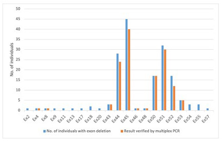
exon deletions analysed by multiplex PCR are depicted in Figure 1. Finally, DNA samples from a total of
135 patients with various single exon deletions were evaluated by the multiplex PCR technique. Amongst
these, exon 45 followed by exon 51 were the commonest single exons found to be deleted in DMD/BMD
patients.
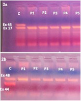
Amongst the evaluated samples, we did not find any discrepancy between the results of MLPA and multiplex PCR for
all the evaluated samples confirming the true positivity of MLPA results in our cohort (Figure 2a & 2b). The SNPs
within 8bp from the ligation sites on probes of the exons and the frequency of alternate alleles in South Asian population
were obtained from gnomAD, ALFA project and the recently published Indian database of 1455 individuals. These
details are represented in Table 1 for all the exons where single exon deletion was identified by MLPA. The
alternate allele frequency of all these SNPs was zero in the South Asian population including the Indian
database.
Figure 1: Total number of individuals with various single exon deletions identified by MLPA in our cohort versus
those which were verified by multiplex PCR.
5 Discussion
Deletions and duplications of the dystrophin gene are the most frequently observed molecular events in DMD/BMD. In
our study, single exon deletions constituted 20.4% of overall deletions/duplication events. Exon 45 followed by 51
was the frequent single exon deletion observed. These findings were similar to previous studies from India
(Basumatary et al., 2013; Deepha et al., 2017) . In this study, we obtained 100% concordant results between
MLPA and multiplex PCR technique for single exon deletion. Analysis of the alternate allele frequency
for SNPs within 8bp of binding site on probes suggested absence of these alternate alleles in South Asian
population.
Amongst various molecular techniques available to detect copy number variations (CNVs), MLPA offers important
advantages as it can be used to identify female carriers, detect duplications and to map deletion and duplication borders
which has important implications for prognosis and management. (Janssen et al., 2005). However, one of the frequent
pitfalls of MLPA reported in literature (Kim et al., 2016) is false deletion results by sequence variations on probe-binding
sites especially for single exon deletions. According to the MLPA design protocol, sequence variations within 8bp
from ligation sites can affect the hybridization or ligation of the MLPA probe (MRC Holland, Amsterdam,
Netherlands).
Figure 2: Multiplex PCR products run on gel electrophoresis. 2a: Top row showing band of exon 45 product and bottom row showing band of exon 17 product (internal control
band).1st lane showing presence of both bands exon 45 and 17 in a control individual and rest of the lanes shows
absence of band for exon 45 only, suggestive of exon 45 deletion in DMD patients. 2b: Top row showing band of exon 48 product (internal control band) and bottom row showing band of exon
44 product.1st lane showing presence of both bands exon 44 and 48 in a control individual and rest of the lanes
shows absence of band for exon 44 only, suggestive of exon 44 deletion in DMD patients.
Also, CA mismatch between the 3’ end of left probe oligonucleotide and the target is found to be associated with the
lowest probe signal among single-nucleotide mismatches and studies have postulated the reason to presence of a single
hydrogen bond for C and A nucleotide (Janssen et al., 2005; Aboul-ela et al., 1985). While efforts are continuously being
taken to prevent designing of probes in those regions, CA mismatch due to new mutagenesis cannot be
prevented and hence interpretation of single exon deletions by MLPA have to be considered always with
caution.
In our study, we did not observe any false positive results for single exon deletions detected by MLPA and multiplex
PCR confirmed the presence of these single exon deletions. Next, we enlisted the SNPs especially those causing CA
mismatches on binding regions of probes within 8bp from ligation sites by obtaining data from dbSNP. The databases like
ALFA project, gnomAD and the recently published Indian database of 1455 samples were analysed for determining the
frequency of alternate alleles. While considerable number of SNPs including those causing CA mismatches especially
between the 3’ end of left probe oligonucleotide and the target were identified, their allele frequency was zero in South
Asian population. This probably explains the concordance of single exon deletion results by MLPA and multiplex PCR.
A previous study in Korean patients has detected a false positive rate of 14.7% (11/75) for single exon
deletions identified by MLPA and factors like CA mismatches in the binding region of the probe and probe
melting temperature (Tm) ≤ 75∘C were found to be causative for false positive MLPA results (Kim et al.,
2016).
Various studies have proven that novel sequence variants (especially C to T transition) account for a considerable
portion of point mutations for DMD gene and hence the probability of new CA mismatches can increase
(Buzin et al., 2005). One solution to partially solve this issue is to upgrade the versions of MLPA regularly
by designing new probes based on the occurrence of new SNPs. Another easier option is to cross check
the results of the single exon deletion detected by MLPA with a simple alternate technique like multiplex
PCR.
6 Conclusion
To conclude, this is the first study in literature that exclusively validated the results of MLPA-detected single exon
deletions of the dystrophin gene by the multiplex PCR technique. Absence of false-positive results for single exon deletion
by MLPA could probably be due to zero frequency of alternate alleles for SNPs. Confirmation of the results of single exon
deletions obtained from MLPA by an alternate technique is however still warranted due to emerging novel single
nucleotide polymorphisms.
References
1. Aboul-ela F, et al. Base-base mismatches. Thermodynamics of double helix formation for dCA3XA3G+
dCT3YT3G (X, Y=A,C,G,T). Nucleic Acids Res. 1985; 13: 4811-4824.
2. Barzegar M, et al. Exon deletion pattern in Duchenne muscular dystrophy in north west of Iran. Iran J
Child Neurol Winter. 2015; 9: 42-48.
3. Basumatary LJ, et al. Deletion pattern in the dystrophin gene in Duchenne muscular dystrophy patients
in northeast India. J Neurosci Rural Pract. 2013; 4: 227-229.
4. Buzin CH, et al. Mutation rates in the dystrophin gene: a hotspot of mutation at a CpG dinucleotide.
Hum Mutat. 2005, 25:177-188.
5. Deepha S, et al. MLPA identification of dystrophin mutations and in silico evaluation of the predicted
protein in dystrophinopathy cases from India. BMC Med Genet. 2017; 18: 67.
6. Flanigan KM, et al. Mutational Spectrum of DMD Mutations in Dystrophinopathy Patients: Application
of Modern Diagnostic Techniques to a Large Cohort. Hum Mutat. 2009; 30: 1657–1666.
7. Janssen B, et al. MLPA analysis for the detection of deletions, duplications and complex rearrangements
in the dystrophin gene: potential and pitfalls. Neurogenetics. 2005; 6: 29-35
8. Kausthubham N, et al. A dataset of variants derived from 1455 clinical and research exomes is efficient in
variant prioritization for early-onset monogenic disorders in Indians. Hum Mutat. 2021; 42: e15-e61.
9. Kim MJ, et al. Pitfalls of Multiple Ligation-Dependent Probe Amplifications in Detecting DMD Exon
Deletions or Duplications. J Mol Diagn. 2016; 18: 253-259.
10. Wang X, et al. Similarity of DMD gene deletion and duplication in the Chinese patients compared to
global populations. Behav Brain Funct. 2008; 4: 20.
Table 1: SNPs within 8 bp from ligation sites on probes and their allele frequency in the South Asian population
for the detected exons from gnomAD, ALFA project and in-house exome data of 727 samples.
a: ALFA project, b: gnomAD, NA: frequency data not available, - indicates absence of SNPs within 8bp from ligation siteson MLPA probes, # indicates those MLPA-detected exon deletions that have been confirmed by multiplexPCR.